Team Medical has been developing
technology to measure cardiac performance
including the real time volumetric flow rate
of the blood being pumped by the heart.
It is estimated that over 500 people die every day from undetected heart ailments because a fast, accurate, and easy means of measuring cardiac pumping function does not exist.
A precision easy-to-use instrument for retail clinics and examination rooms and hospital floors is one product concept using this innovative technology
Save lives by making cardiac function health measurements routine for everyone. Make early diagnosis routine, such as during pregnancy check-ups.
Today, no one can know how well a heart is actually pumping blood for a simple reason: no means exists for measuring the rate that blood flows out of the left ventricle.
Team medical has been developing technology to measure blood flow rate out of the left ventricle and evaluate cardiac function.
The development goals have been to:
•
measure in real time the rate at which blood flows from the heart using noninvasive means,
including ultrasonic Doppler, and
•
measure cardiac health such as by determining cardiac power and cardiac output during
each heartbeat.
Non-invasive measurements to be accurate, automatic, fast, easily charted, and cost-effective outside of specialized labs

The goal is non-invasive measurements in an office or clinic to make routine accurate measurements that cost much less than echocardiograms and other tests.
Team Medical has U.S. patents to meet both of these goals with other patents pending.
The patents address two important problems.
•
The patents describe a new means to determine noninvasively the volumetric flow rate of blood.
•
The patents describe a novel signal processing technique that overcomes problems inherent in
traditional analysis of time-varying signals, such as those occurring when measuring blood flow rates.
Together the patents present solutions to the two fundamental problems associated with noninvasively measuring in real time the volumetric flow rate of blood.
1.
What is the cross sectional area of the blood vessel?
Doppler methods can provide the velocity of flowing blood, but unless the cross sectional area of the blood vessel, such as the ascending aorta, is known the volumetric flow rate cannot be calculated.Previous approaches to estimate the cross-sectional area have attempted to image the blood vessel or bounce signals off the side walls. These methods suffer from many deficiencies, such as needing to get an ultrasonic probe very close to the blood vessel.
Team Medical's method is completely new.
It recognizes that the rate of change of the blood flow rate itself provides information on the dimensions of the blood vessel and uses this information to determine the dimensions of the blood vessel.
An additional benefit is that other clinically relevant information is produced, such as a measure of the elasticity of the ascending aorta.

This patented approach also describes how to avoid problems that can confound using Doppler ultrasound.
In particular, the patents describe how to discriminate automatically between the signal that comes from the blood flowing in the vessel from the part of the signal that comes back from all of the other slow moving and stationary tissue structures.
2.
What is the frequency spectrum of the returned signal when the flow rate is changing and the signal has been quantized by the measurement process?
The velocity profile across a blood vessel changes during the cardiac cycle.For major arteries this change is significant and for the aorta the blood flow rate changes quickly enough that the flow rate is never steady state. Consequently, the signals emanating from such sources also change constantly, which means that the spectral content of the signals also changes constantly.
Traditional methods of spectral analysis that rely on Fourier and other transform methods cannot handle such non-stationary signals when high resolution is needed. Traditional methods cannot obtain a long enough data stream to provide good frequency resolution.
Methods based on analyzing time-series, such as AR and ARMA methods, are poorly suited. For example, the spectra calculated using time series methods strongly depend on the order of the model selected, which cannot be reliably determined.
Further compounding the problem is that the data available have been distorted by the measurement process. The measurement system converts the continuous analog signal into numeric digital values. The analog to digital conversion process puts measured values into bins, a process that introduces digitization errors.
Team Medical's patented approach avoids these problems.
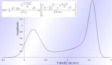
Team Medical's novel approach calculates the complete continuous range of velocities across the inside of the blood vessel (using continuous spectral density functions of frequency) using measurements from the time-varying (non-stationary) signals even though the measured values have digitization errors.
Among the features of the Team Medical method are a novel regularization method that leads to unique solutions (to remove the uncertainty caused by the digitization error) and that does not require using external weighting factors, overcoming a deficiency of methods such as Tikhonov regularization.
Team Medical developed technical approaches to overcome problems encountered when obtaining data in the real world.
For example, any interrogation using an output signal, such as an ultrasonic beam, will get signals back from not only the region of interest where blood is flowing, but also from many other tissues.
It is impractical to expect a clinically useful device to employ a very narrow beam that only interrogates a tiny hard to locate region deep inside a patient.
The patented Team Medical method for determining channel dimensions has been designed to automatically sort through the returned signal and discriminate between the region that has the blood flow and all of the other regions.
A variety of possible implementations exist. In practice, the overall process consists of three steps:
1.
Send a signal into the tissue and get back a reflected signal.
2.
Process the reflected signal to get the distribution of velocities
of the tissues and determine the area of the channel through which blood is flowing.
3.
Use the distribution of velocities to calculate the average flow
rate of the blood and multiply this value times the area of the channel to obtain the blood's volumetric flow rate.
The volumetric flow rate obtained using the Team Medical method is the much more valuable instantaneous flow rate, not a long term average flow rate such as is measured with cardiac catheters.
In somewhat more detail, the following steps are used:
1.
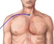
Using an interrogating signal, such as ultrasonic Doppler,
receive a signal back from the region of interest, such as where the ascending aorta carries blood from the heart.
2.
Using the signal processing method calculate the signal's spectral density function.
3.
Calculate the velocity spectrum from the signal's spectral density.
4.
Calculate the cross sectional area of the channel, such as the ascending aorta,
based on changes in the velocity spectrum every, for example, 5/1000 ths of a second.
5.
Calculate, using the velocity spectrum, the average velocity.
Use this value and the cross sectional area to calculate the volumetric flow rate.
6.
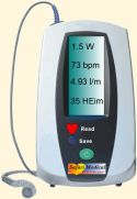
Display the volumetric flow rate directly or use various derived parameters,
such as the rate of change in velocity (blood flow acceleration) to evaluate the condition of the patient's heart.
These data can be used in other ways, too. For example, the instantaneous volumetric flow rate data can be used in conjunction
with an EKG to show heart pumping activity along with the myocardium's electrical activity.
Multiple United States patents have been issued for the Team Medical cardiac function measuring method.
Click on "Patents" in the main menu near the top of this page for a complete list of them. Other patent applications are pending.
Below are links to two representative patents.
•
United States Patent 8,784,327 for
"Method and system for obtaining dimension related information for a flow channel"
US8784327.PDF (click to download)•
United States Patent 7,806,825 for
"Diagnostic signal processing method and system"
US7806825.PDF (click to download)
The technology described differs from all other cardiac output measurements known to Team Medical. For additional information regarding these technologies and
opportunities to license them or participate in their development, please contact us.